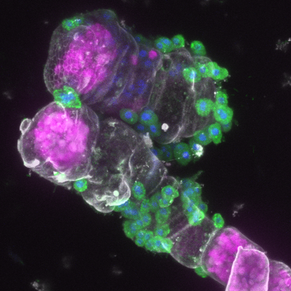
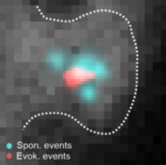
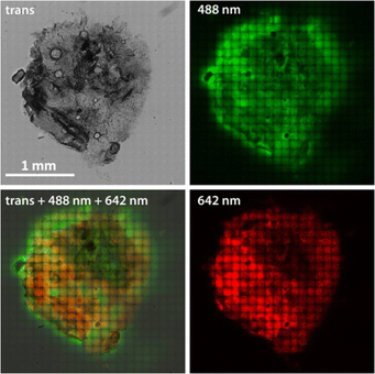
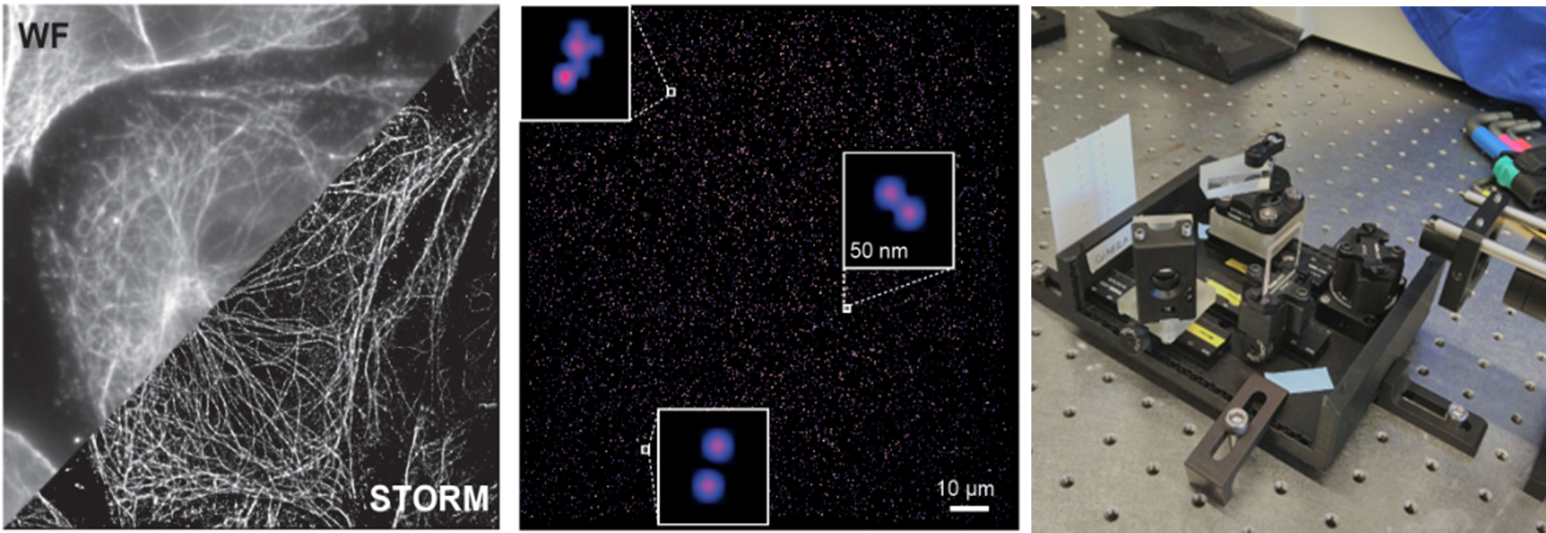
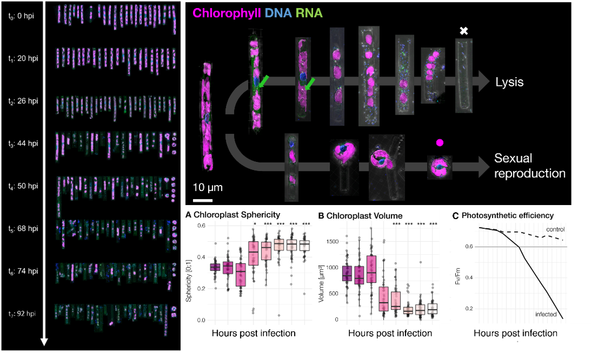
Subcellular marine microscopy

I am Marie Walde, a biophysicist studying small eukaryotes in the marine plankton with advanced 3D light microscopy and image analysis at the marine reserach station in Roscoff/Bretagne (France).

My research is funded by the German Research Council (Deutsche Forschungsgemeinschaft)

SEE MORE

About Me

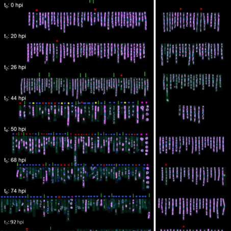
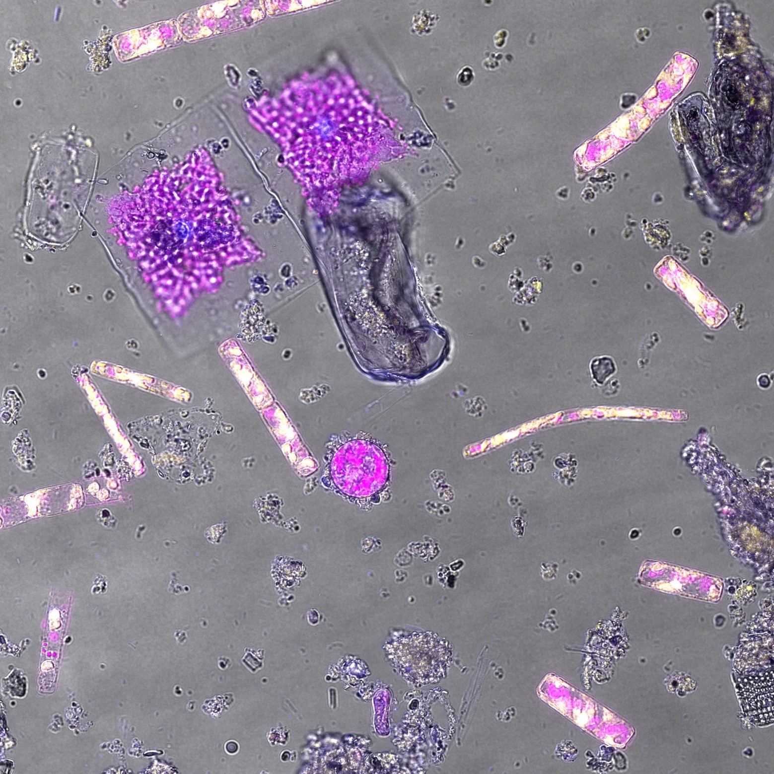
I work on deciphering the morphologies and cellular interactions of unicellular marine microbes, in particular understudied small protists.
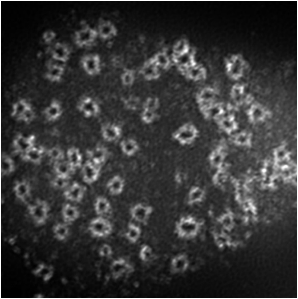
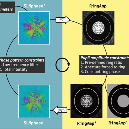
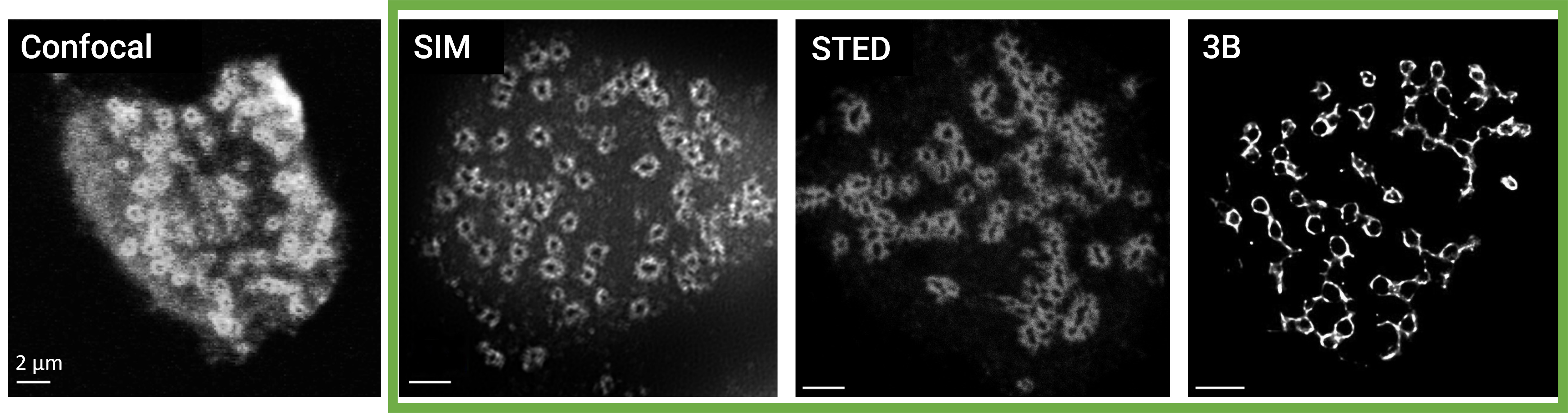
Towards this goal, I develop and use tailored bioimaging and image processing methods, including automated and super-resolution microscopy to address their challenging diversity and connect information across large scale ranges.

Career

Postdoctoral Fellow

Station Biologique de Roscoff, France September 2021 – Present

Walter Benjamin Fellowship by the German Reserach Foundation (DFG)

Project SymbioScope: Assessing the ecological diversity and dynamics of symbiotic interactions in a key phytoplankton by automated high-content 3D microscopy

Visiting Scientist

Uppsala Universitet, Sweden July 2021 – August 2021

Hosted by Prof. Dr. Fabien Burki, Department of Organismal Biology
Automated microscopy to elucidate unknown eukaryotic diversity

Postdoctoral Fellow

Station Biologique de Roscoff, France December 2019 – June 2021

Stratégie d'attractivité durable (SAD) International Postdoctoral Fellowship by the Région Bretagne: Competitive fellowship aimed at international researchers who bring new methods and expertise to research establishments based in Bretagne

Supervised by Prof. Dr. Nathalie Simon & Dr. Colomban de Vargas,
UMR 7144 Adaptation and Diversity in Marine Environment,
CNRS/Sorbonne Université, France
Project IMASTAN: Automated imaging of phytoplankton in the SOMLIT-Astan time series

Imaging Scientist

Max Delbrück Center, Germany Januar 2017 – November 2019

Systems Biology Imaging Platform, headed by Andrew Woehler, PhD
Max Delbrück Center (MDC) for Molecular Medicine in the Helmholtz Association

Superresolution fluorescence neuroimaging

Education

PhD in Biomedical Imaging & Microscopy

Friedrich-Schiller-Universität Jena, Germany September 2017

Leibniz Institute of Photonic Technologies & Institute of Physical Chemistry
Supervised by Prof. Dr. R. Heintzmann, Nanobiophotonics Research Group
Research Project: Development of a holoscopic imaging system and applied high-resolution fluorescence microscopy

Master of Research in Molecular Biophysics

King's College London, UK September 2010

Randall Centre for Cell & Molecular Biophysics
Research Project: High-resolution fluorescence microscopy with photoswitchable proteins

Bachelor of Science in Biophysics & Physics

Humboldt-Universität zu Berlin, Germany September 2009

Research Project: Resolution improvement with structured illumination fluorescence microscopy applied to biological research

Experimental Methods

Fluorescence microscopy, automated microscopy, super-resolution microscopy, image processing & quantitative analysis, plankton sampling and filtration, plankton culturing, optical design & calibration, instrument control, fluorescent labelling & immunocytochemistry, in-situ hybridization, interferometry, 3D modelling and printing

Research

Publications

  • M. Walde, C. Camplong, C. de Vargas, A.-C. Baudoux & N. Simon (2023). Viral infection impacts the 3D subcellular structure of the abundant marine diatom Guinardia delicatula. Frontiers in Marine Science 9, 1034235.
    DOI: 10.3389/fmars.2022.1034235

  • M. M. Sandin & M. Walde (2022). oligoN-design: First beta version. GitHub Releases.
    Bioinformatic pipeline for the high-throughput design of specific probes for fluorescence in situ hybridization of only genetically known organisms.
    DOI: 10.5281/zenodo.7473220.

  • Z. Farsi, M. Walde, A. E. Klementowicz, F. Paraskevopoulou & A. Woehler (2021). Single synapse glutamate imaging reveals multiple levels of release mode regulation in mammalian synapses. iScience, 24(1), 101909.
    DOI: 10.1016/j.isci.2020.101909

  • M. Walde & A. Woehler (2018). Spectral STORM - Multi-colour super-resolution imaging with a single excitation laser. European Light Microscopy Initiative (ELMI2018), Dublin.
    DOI: 10.5281/zenodo.3833716

  • M. Walde, A. Jost, K. Wicker & R. Heintzmann (2017). Engineering an achromatic Bessel beam using a phase-only spatial light modulator and an iterative Fourier transformation algorithm. Optics Communications, 383, 64–68.
    DOI: 10.1016/j.optcom.2016.08.050

  • M. Walde (2017): Development of a holoscopic imaging system and applied high-resolution fluorescence microscopy. Friedrich-Schiller-Universität Jena. PDF

  • Badgaa, R. Büchler, N. Wielsch, M. Walde, R. Heintzmann, Y. Pauchet, A. Svatos, K. Ploss & W. Boland (2015). The Green Gut: Chlorophyll Degradation in the Gut of Spodoptera littoralis. Journal of Chemical Ecology, 41(11), 965–974.
    DOI :10.1007/s10886-015-0636-0 PDF

  • M. Walde, J. Monypenny, R. Heintzmann, G.E. Jones & S. Cox (2014). Vinculin binding angle in podosomes revealed by high resolution microscopy. PLoS ONE, 9(2).
    DOI: 10.1371/journal.pone.0088251

  • K. Wicker, M. Walde, E. Oldewurtel, L. Hirvonen, O Mandula, S. Sindbert & R. Heintzmann (2010). Structured Illumination and Image Inversion Interferometry. Biophysical Journal, 98(3), 619a.

Say Hello

Ecology of Marine Plankton
Station Biologique de Roscoff
CNRS & Sorbonne Université
Place Georges Tessier, 29680 Roscoff, FRANCE

Email

marie.walde@sb-roscoff.fr

Other Platforms